Anatomical Models
High-fidelity medical models engineered for educational demonstration, surgical planning, patient consultation, and veterinary diagnostics.
Primary Applications
Medical Universities Clinical Consultation Surgical Simulation Veterinary Schools Biomedical Exhibitions
Key Features
Life-Size Accuracy Magnetic Articulation Pathology Overlays Cross-Sectional Views Durable PVC Casts
Single Tap to Scroll • Double Tap to Open Catalog
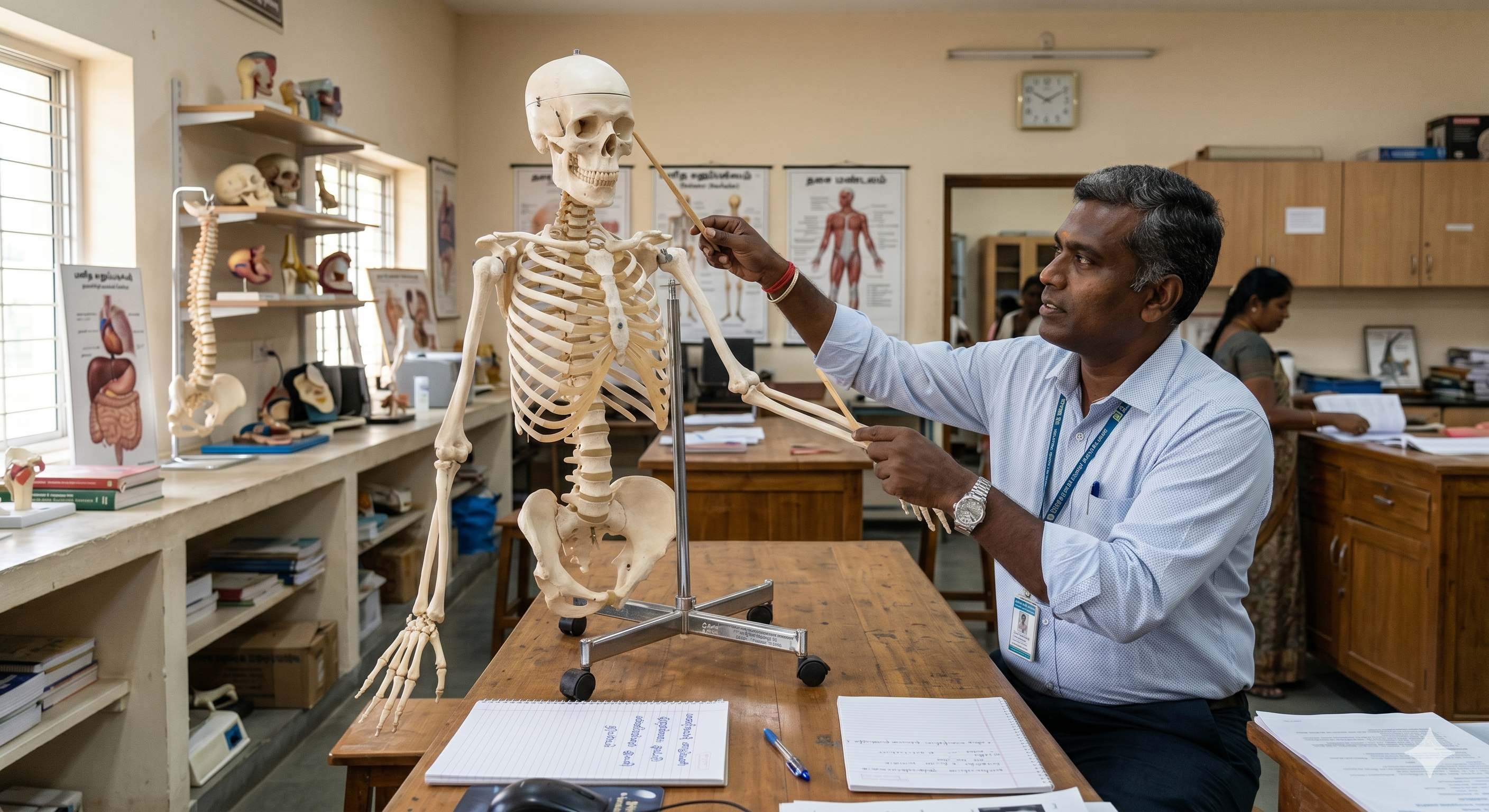
Human Skeleton Models
- Life-size and miniature articulated skeletal systems.
- Shows muscle origins and insertions with color-coded markers.
- Features herniated discs and flexible spinal columns.
- Mounted on heavy-duty caster stands for easy classroom mobility.
Access Catalog
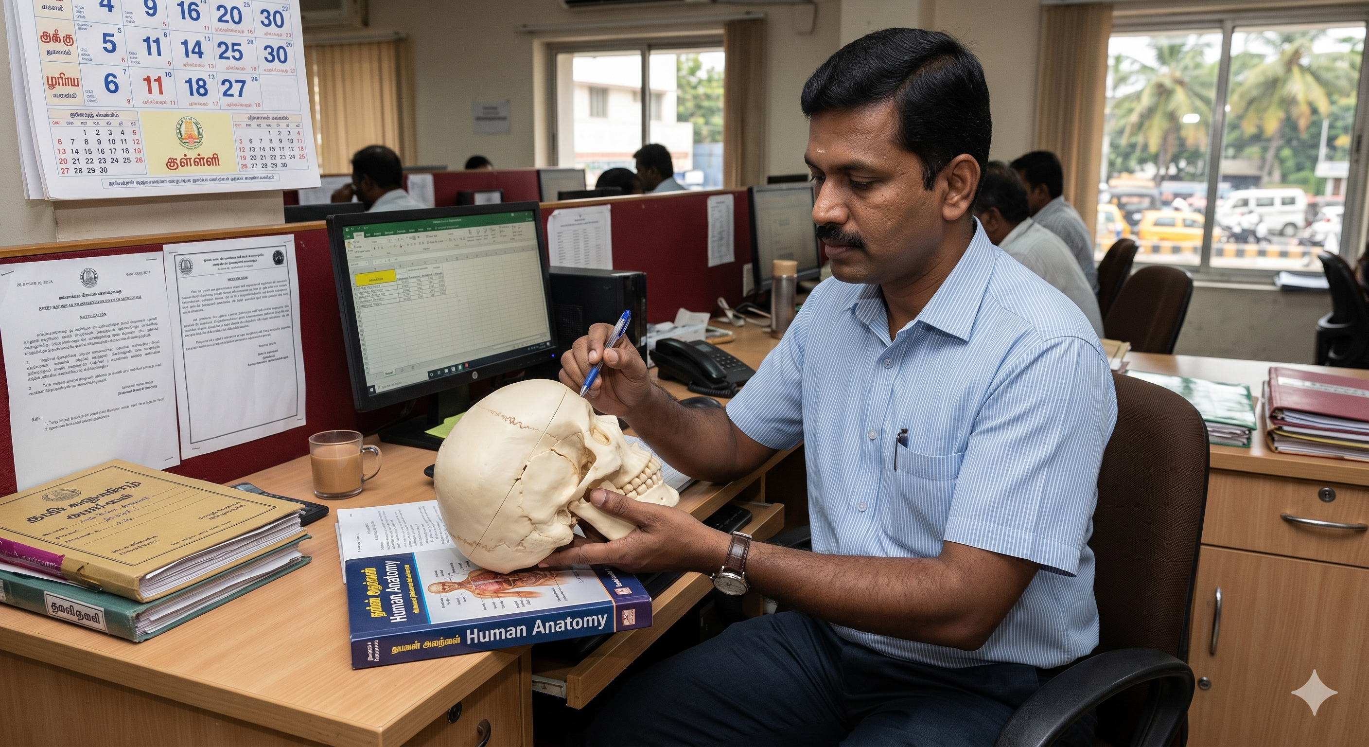
Skull Models
- Dissectible 3-part skulls (calvarium, base, and mandible).
- Exploded Beauchene skulls mapping all 22 individual bones.
- Transparent models revealing sinus cavities and tooth roots.
- Detailed cranial sutures and foramina for neurology studies.
Access Catalog
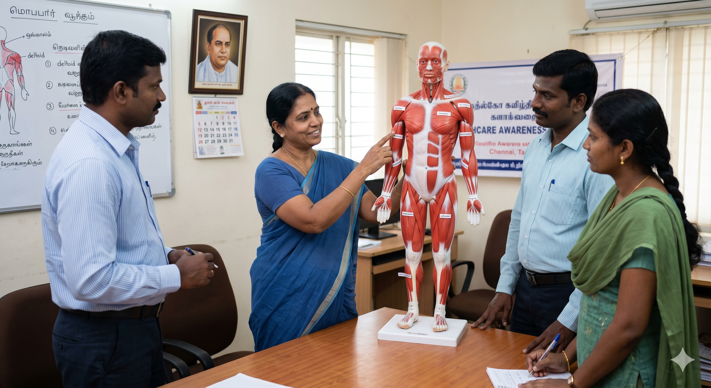
Muscular System Models
- Full-body muscular figures detailing superficial and deep muscles.
- Removable muscle groups exposing underlying vascular structures.
- Includes specific models for arms, legs, and facial musculature.
- Essential for sports medicine, physical therapy, and kinesiology.
Access Catalog

Nervous System Models
- Relief boards mapping the entire central and peripheral systems.
- Spinal cord cross-sections showing nerve root branchings.
- Autonomic nervous system boards detailing sympathetic pathways.
- Critical for chiropractic and neurology educational programs.
Access Catalog
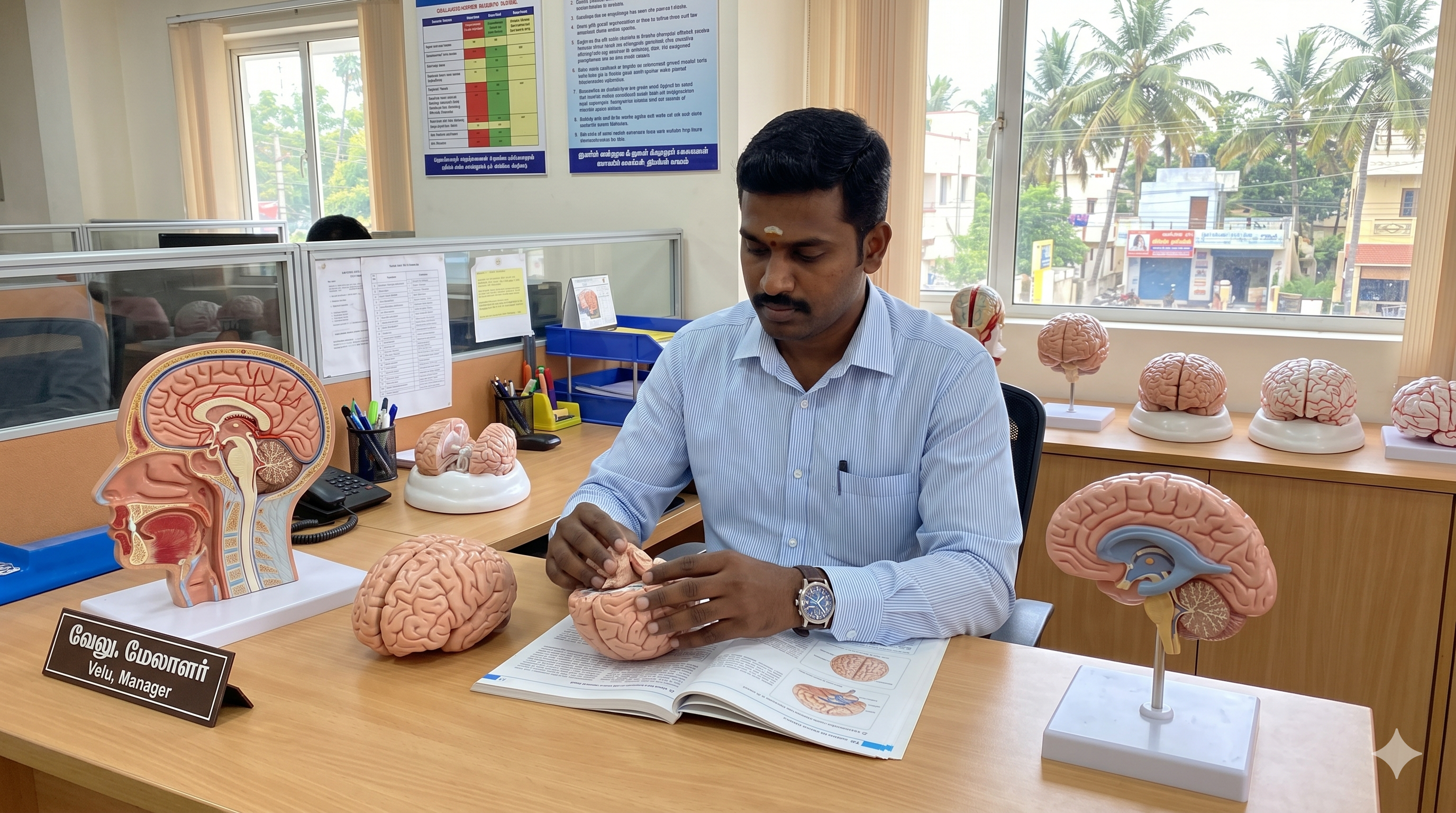
Brain Models
- Magnetically dissectible models separating into 2 to 9 parts.
- Color-coded mapping of functional regions (motor, sensory, visual).
- Reveals the brain stem, cerebellum, and intricate circle of Willis.
- Fits securely within matching human skull models for context.
Access Catalog
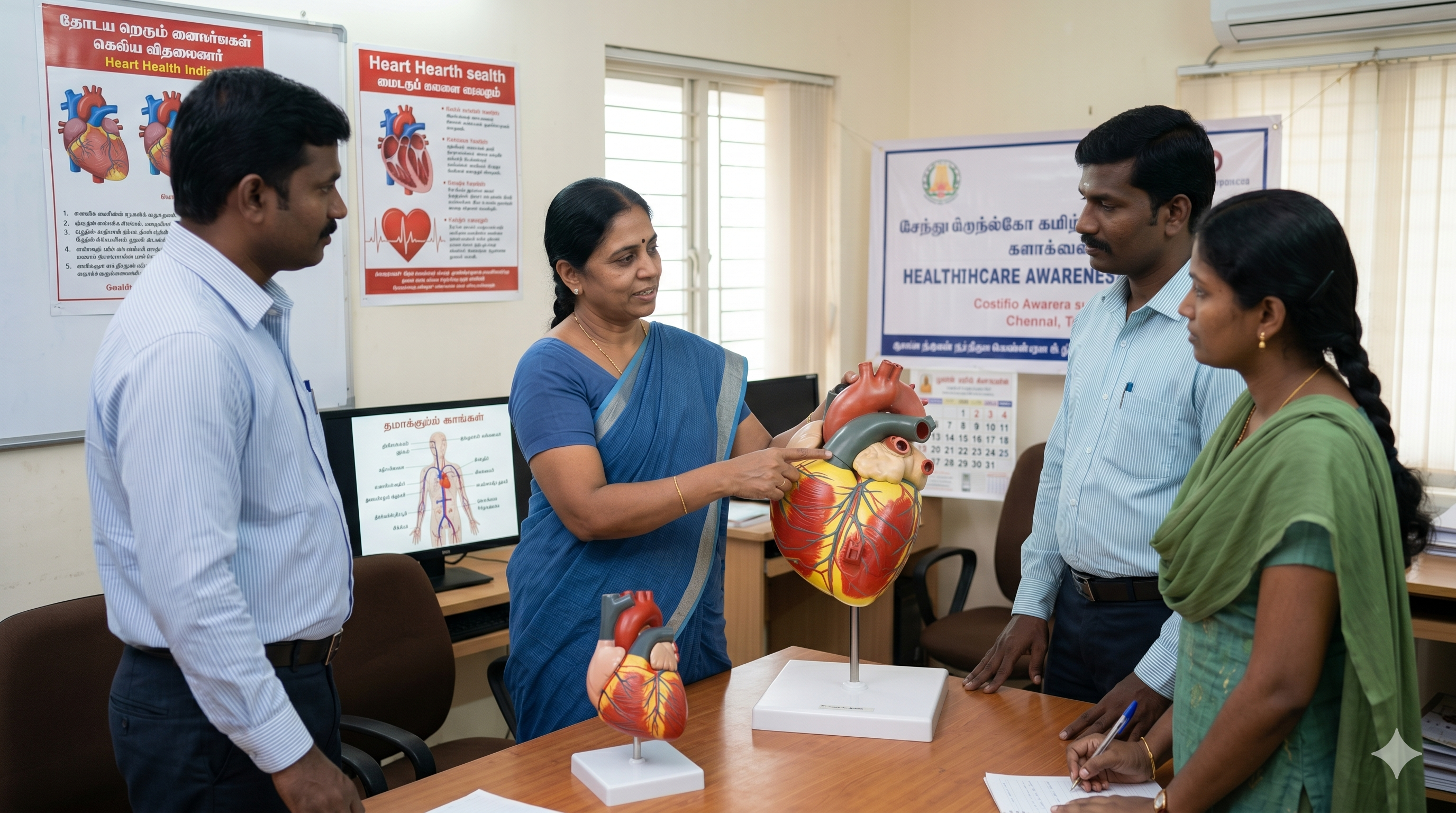
Heart Models
- Giant 2x to 5x life-size models for lecture hall visibility.
- Removable anterior walls revealing atria, ventricles, and valves.
- Shows coronary bypass vessels, pacemakers, and conductive tissues.
- Crucial for cardiology patient consultation and pre-op explanations.
Access Catalog
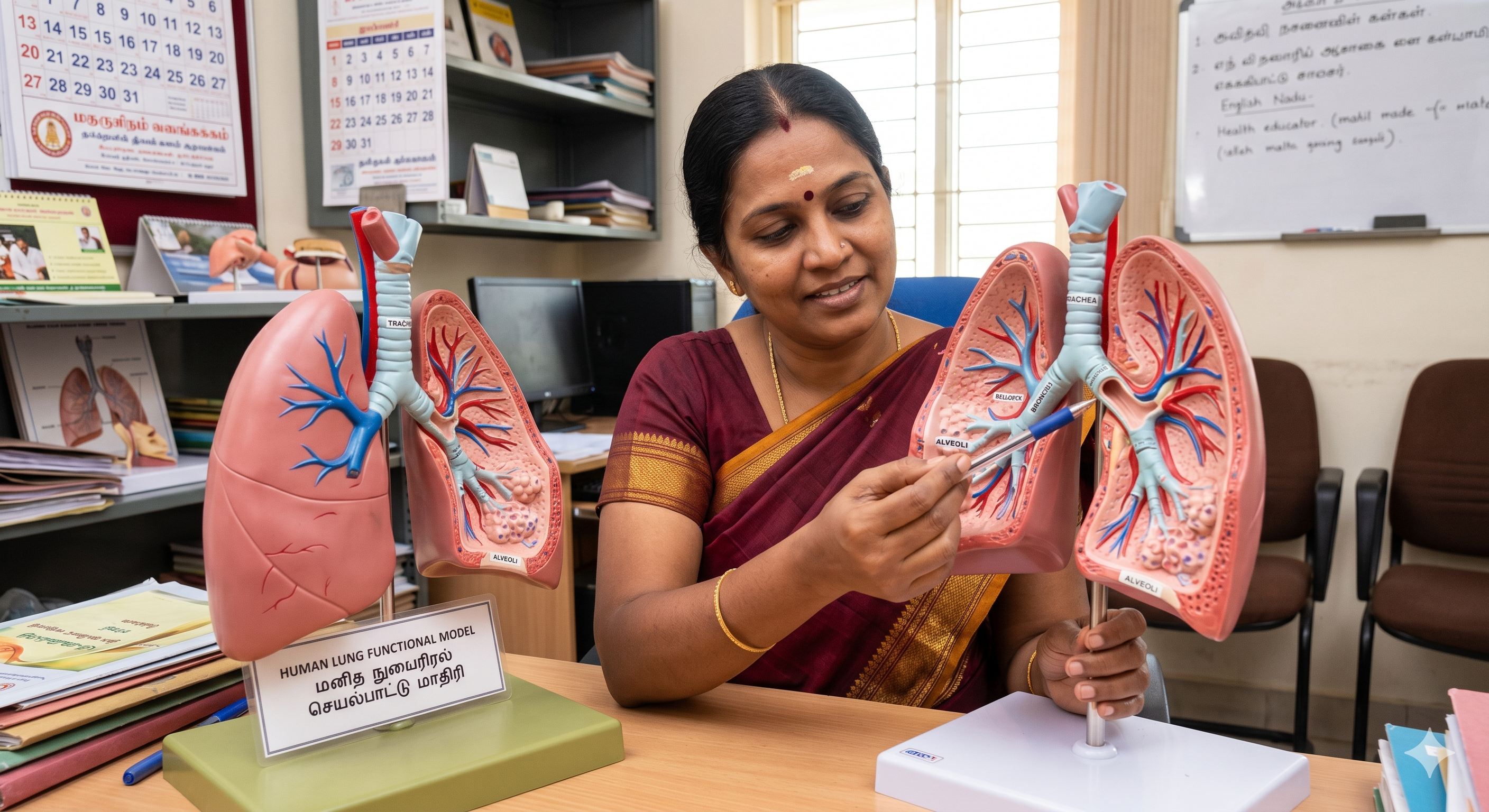
Lung Models
- Bronchial tree models mapping the respiratory tract subdivisions.
- Transparent lung casts showing the pulmonary vascular system.
- Segmented models demonstrating the individual bronchopulmonary lobes.
- Includes models depicting the diaphragm and respiratory mechanics.
Access Catalog
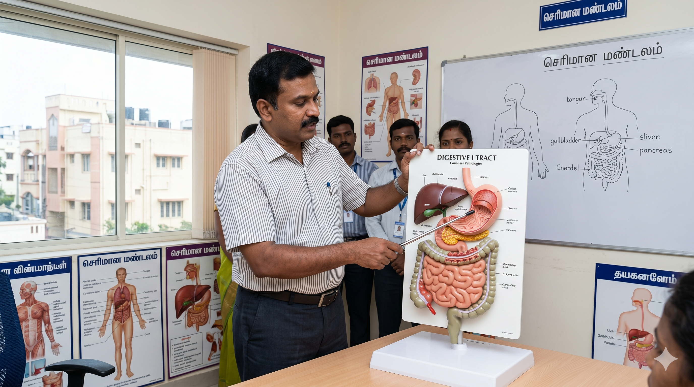
Digestive System Models
- Relief models showing the entire GI tract from mouth to rectum.
- Removable stomach halves revealing internal mucosal folds (rugae).
- Detailed liver/gallbladder models mapping the biliary duct system.
- Intestinal villi models magnified 100x for histology education.
Access Catalog
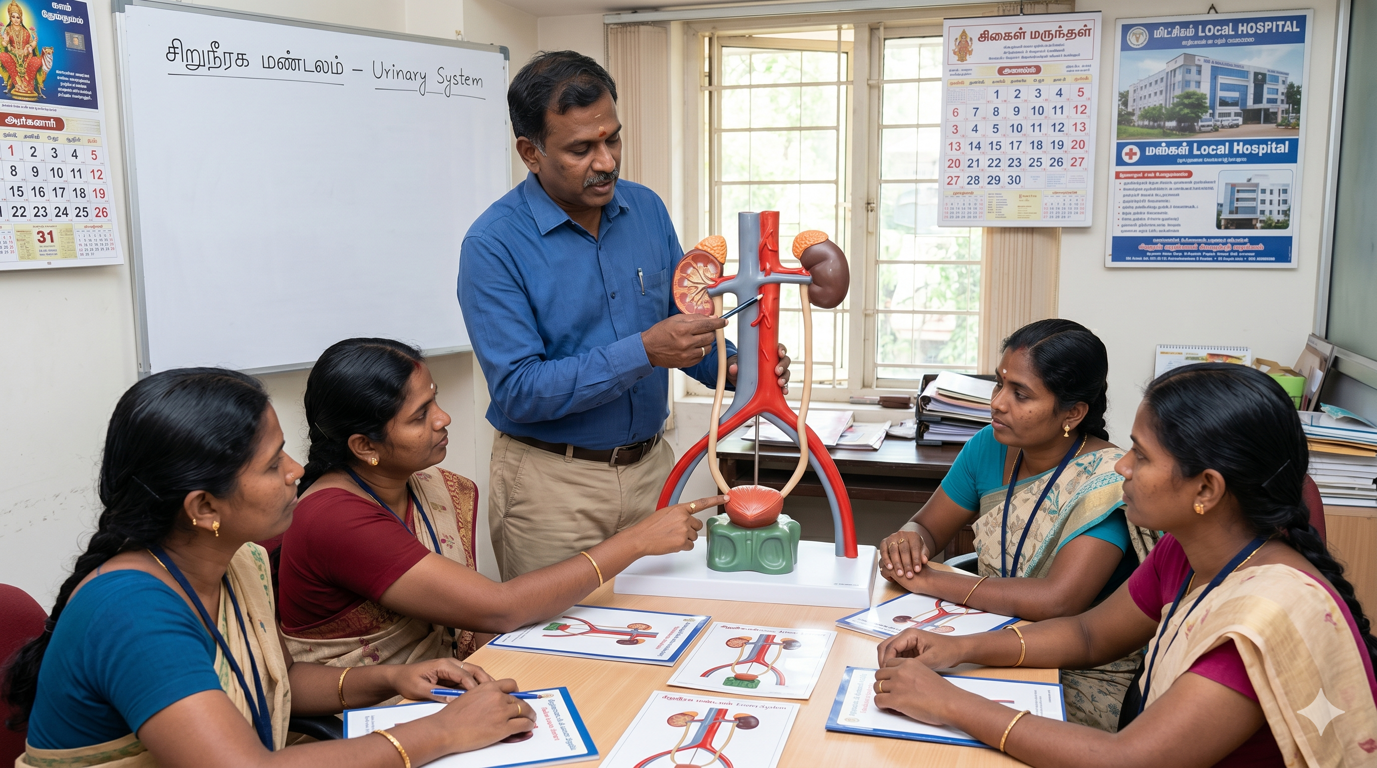
Urinary System Models
- Dual-sex urinary systems detailing kidneys, ureters, and bladder.
- Kidney cross-sections displaying the cortex, medulla, and calyces.
- Magnified nephron and renal corpuscle models for advanced physiology.
- Shows adrenal glands and major abdominal blood vessels.
Access Catalog
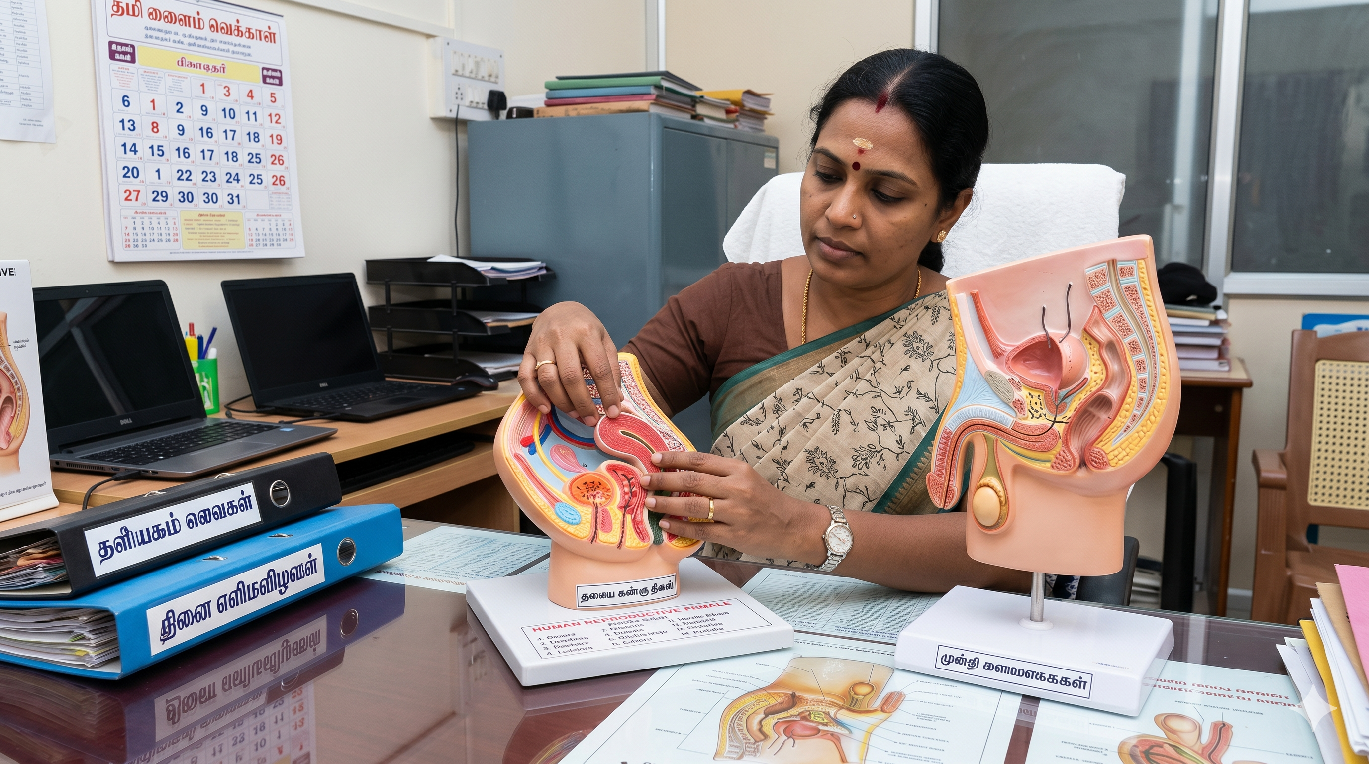
Reproductive System Models
- Median sagittal sections of the male and female pelvis.
- Removable genitalia detailing internal and external structures.
- Pregnancy series models tracking fetal development stages.
- Essential tools for obstetrics, gynecology, and urology clinics.
Access Catalog
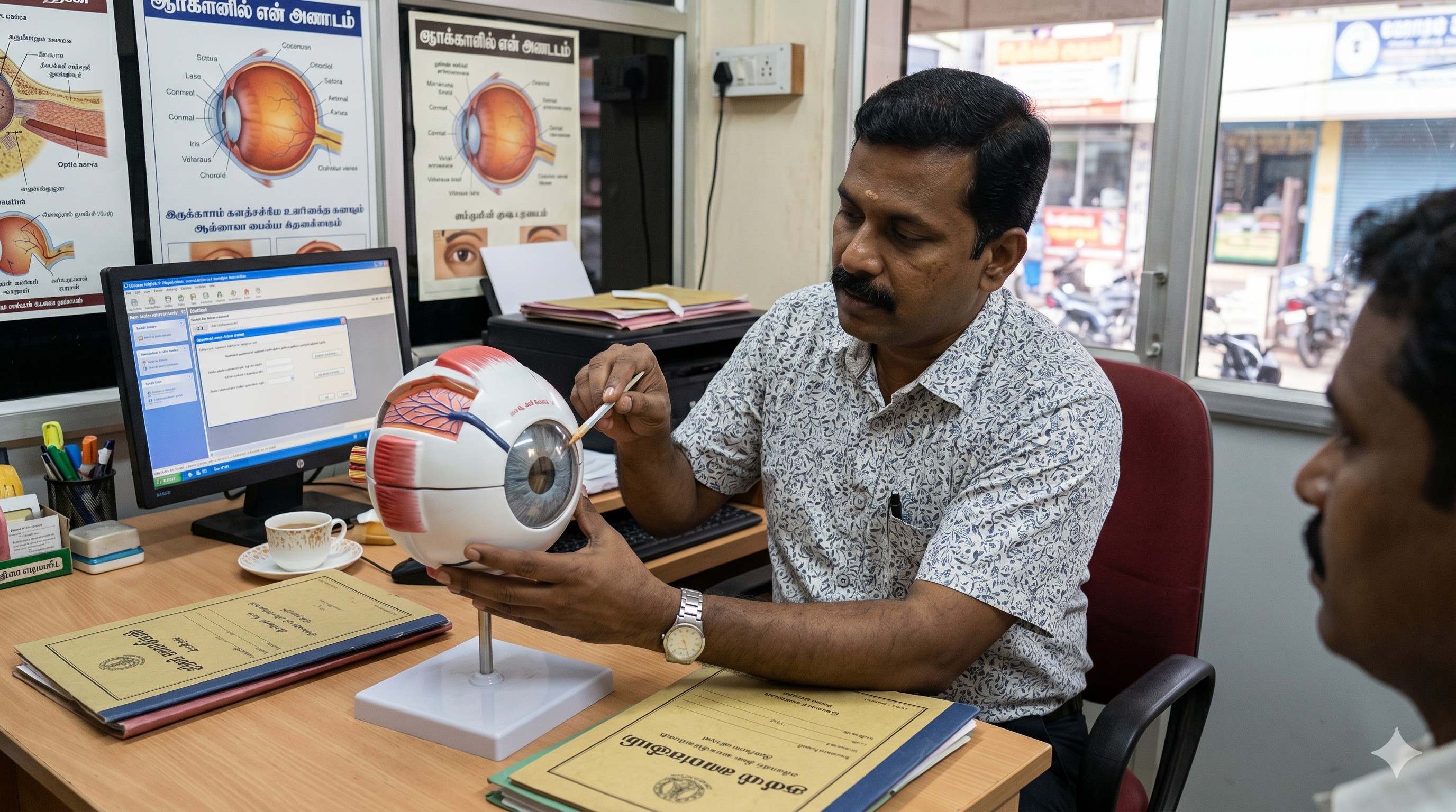
Eye Models
- Giant 3x to 6x models dissectible into multiple layers.
- Reveals the sclera, choroid, retina, and removable vitreous humor.
- Details the extraocular muscles and optic nerve attachments.
- Critical for optometry training and cataract surgery explanations.
Access Catalog
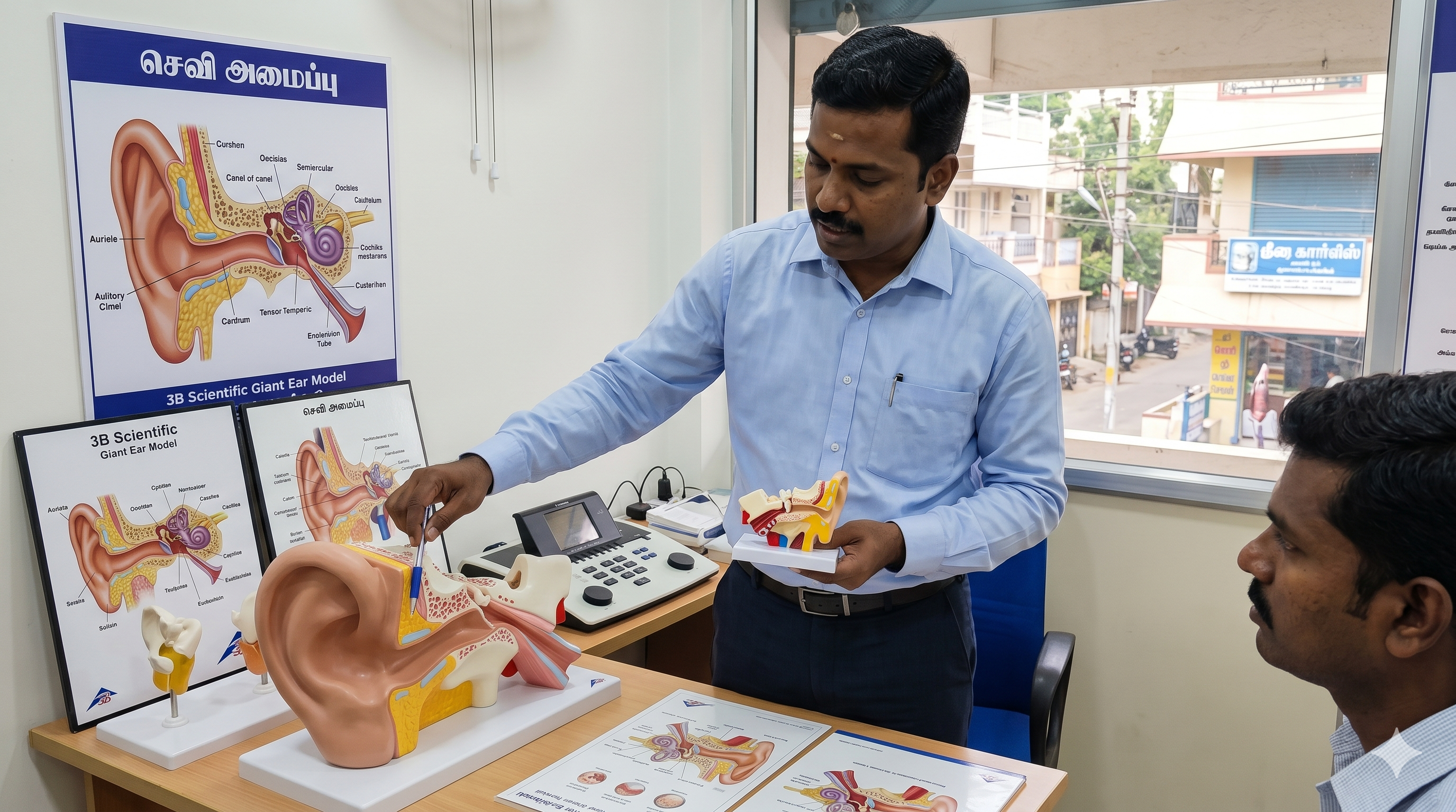
Ear Models
- Enlarged models depicting the outer, middle, and inner ear.
- Removable tympanic membrane with attached auditory ossicles.
- Detailed cochlea and vestibular labyrinth for audiology studies.
- Perfect for explaining hearing loss and cochlear implants.
Access Catalog

Dental / Tooth Models
- Giant molar models dissectible to reveal enamel, dentin, and pulp.
- Full jaw models displaying adult versus deciduous (baby) teeth.
- Orthodontic models equipped with brackets, wires, and elastics.
- Pathology models demonstrating caries, gingivitis, and root canals.
Access Catalog
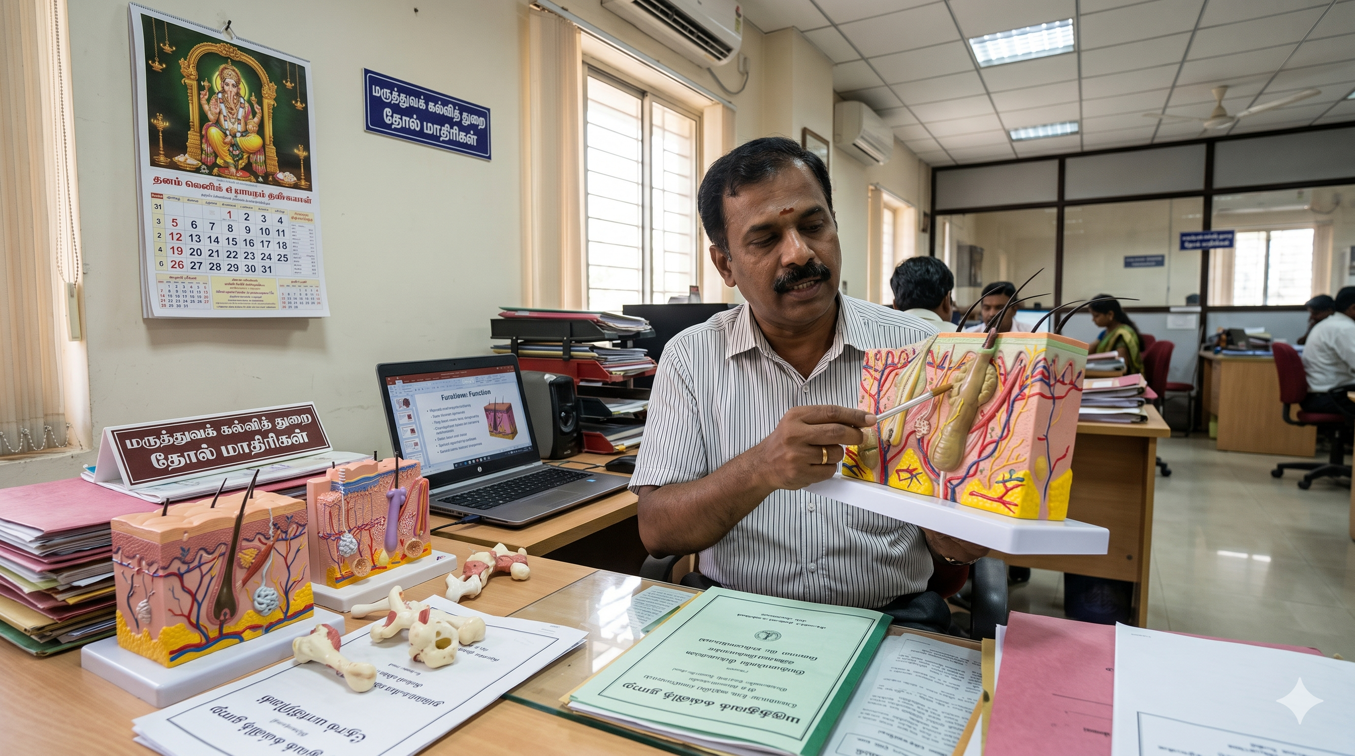
Skin Models
- Highly magnified blocks showing epidermis, dermis, and subcutis.
- Details hair follicles, sebaceous glands, and sweat glands.
- Displays distinct tactile receptors and nerve ending structures.
- Burn and wound pathology models for dermatology classification.
Access Catalog
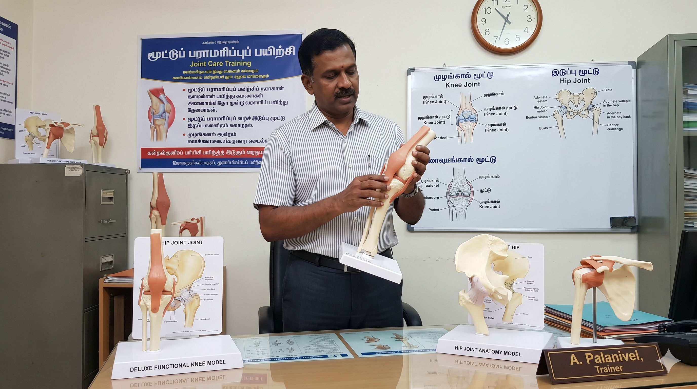
Joint Models (Knee, Hip, etc.)
- Functional models mimicking true biomechanical flexion and extension.
- Features flexible artificial ligaments, tendons, and menisci.
- Highlights common sports injuries (ACL tears, rotator cuff damage).
- Includes models demonstrating osteoarthritis and joint replacement.
Access Catalog

Torso / Full Body Models
- The ultimate classroom centerpiece with up to 40 dissectible parts.
- Dual-sex and sexless variants with interchangeable genital inserts.
- Exposed spinal columns and open backs revealing the spinal cord.
- Provides a comprehensive, unified view of human splanchnology.
Access Catalog

Pathological / Disease Models
- Specialized models demonstrating diseased states versus healthy anatomy.
- Includes fatty livers, smoker's lungs, and atherosclerotic arteries.
- Highlights cancer staging in colons, breasts, and prostates.
- Invaluable for patient education and health awareness campaigns.
Access Catalog
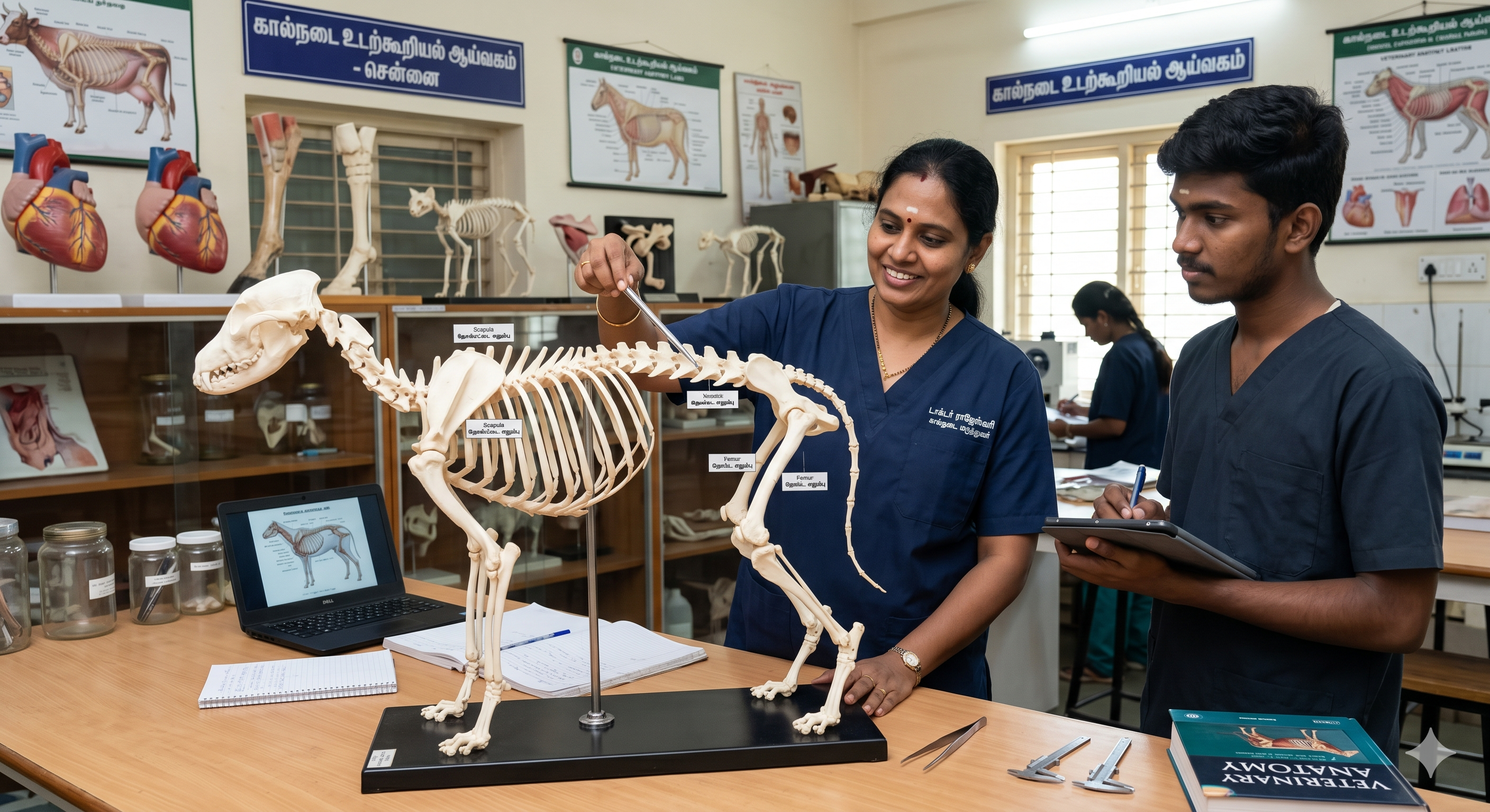
Veterinary Anatomical Models
- Detailed skeletal and organ models for canine, feline, and equine species.
- Life-size animal joints demonstrating patellar luxation and hip dysplasia.
- Dental models mapping domestic animal dentition for veterinary dentistry.
- Crucial for veterinary university training and pet owner consultations.
Access Catalog
No Categories Found
Try adjusting your search terminology.
Clinical Simulation Standards
Anatomical models bridge the gap between flat textbook diagrams and actual surgical environments. Selecting high-fidelity models ensures that medical students develop precise spatial awareness and patients fully comprehend their diagnoses.
Magnetic Dissection Architecture
Enhanced UsabilityModern premium models replace fragile plastic pegs with embedded rare-earth magnets. This allows for rapid, seamless disassembly and reassembly of complex organs like the brain and heart, preventing the wear and tear associated with classroom use.
Hand-Painted Cartography
Fidelity & AccuracyThe highest quality anatomical casts are individually hand-painted by medical artists. This ensures absolute precision when delineating cranial nerve pathways, muscular origins (red) and insertions (blue), and fine vascular networks.
Micro-Anatomical Magnification
Histological InsightCertain physiological processes cannot be taught at life-size. Models that scale up structures by 100x to 10,000x—such as the renal nephron, intestinal villi, or skin layers—are critical for explaining cellular-level filtration and absorption.
Pathological Contrasts
Patient CommunicationModels depicting disease states (e.g., a multi-stage herniated disc or a segmented diseased artery) provide unparalleled value in the clinic. They offer tangible, non-threatening visuals that help patients understand the necessity of a prescribed surgical intervention.
Model Specification Matrix
| Model Category | Primary Educational Value | Key Feature to Specify |
|---|---|---|
| Articulated Skeletons | Osteology, Kinesiology, Orthopedics | Flexible spine vs. wire-mounted rigid spine. |
| Dissectible Torsos | General Splanchnology, Nursing | Open back (exposing spinal cord) vs. closed back. |
| Functional Joints | Sports Medicine, Physiotherapy | Artificial ligaments mimicking true flexion/extension. |
| Neuro-Anatomical (Brain/Skull) | Neurology, Neurosurgery | Magnetic part attachment; mapped Brodmann areas. |
| Pathology Specific | Patient Consultation, Public Health | Side-by-side healthy vs. diseased state comparison. |
| Veterinary Molds | Animal Husbandry, Vet Sciences | Species specific (Canine/Feline) common injury markers. |